

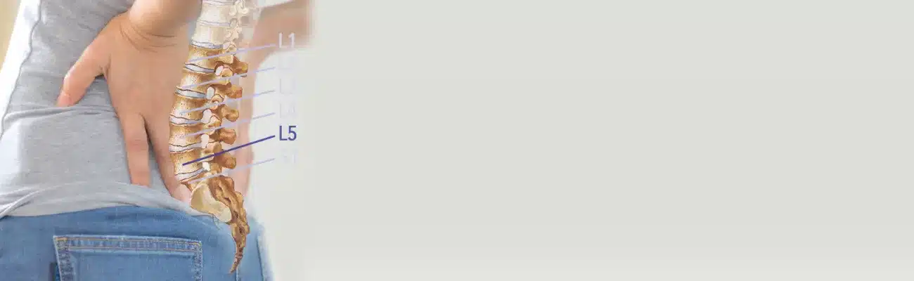
L5-S1 Herniated Disc: Symptoms, Treatment and Recovery Time
An L5-S1 herniated disc or disc bulge often shows up as low back pain or a cramp-like ache in one buttock. When a slipped disc at L5-S1 reaches the S1 nerve root, pain or numbness can run down the leg to the outer foot. Sitting tends to worsen the back pain, and a cough can sharpen the leg pain. The calf can weaken too.
In my experience, most L5-S1 disc damage follows an injury, sudden or repeated, or strain repeated day after day. Long sitting, slouching and lifting in poor positions add to it. After L4-L5, this is where I find disc problems most often.
Care begins with an examination, so that any scan is read against the strength, reflexes and feeling of the leg. Gentle chiropractic care without twisting is combined with physiotherapy and hands-on soft tissue work. Precise exercise follows once the back has settled, always within what the patient can comfortably tolerate. The sections that follow explain healing without surgery, the S1 and L5 nerves, each stage from bulge to fragment, MRI terms, recovery time and surgery.
This guide, L5-S1 Disc Bulge & Herniation: Symptoms, Causes and Care, is published by Chiropractic Specialty Center, Bukit Damansara (KL) main center: WhatsApp+60 17 269 1873 Call+603 2093 1000
L5-S1 Herniated Disc: Key Takeaways
| Point | What to know |
|---|---|
| Where L5-S1 is | The lowest disc of the lower back, between the fifth lumbar vertebra (L5) and the sacrum (S1), the bone at the back of the pelvis. It is one level, with one disc. |
| Recovery time | There is no fixed time. Without improvement by the fifth session, a reassessment is due; with none by the 10th, continuing or stopping is decided together with the chiropractor. |
| Healing without surgery | A damaged disc never returns to its original state, with or without an operation. Surgery is still the last option, and I judge progress by the retested nerve findings. |
| How serious it is | To some degree, always. On my scale of 0 to 10, a true bulge rates 1 or 2. A disc that reaches the S1 or L5 nerve root rates 3 to 5. |
| Which nerve it affects | Usually the S1 root, which crosses the back of the disc: a large disc just off center can press on it (Berry 2019). Material out in the side opening can reach the L5 root. |
| Bulge or herniation | Slipped disc is the everyday name for every stage. A true bulge has swollen without reaching the nerves; in a herniation the soft center has broken through the outer wall. |
| Scan versus symptoms | Bulges are common without pain: in an MRI study of 98 adults with no back pain, 52 percent had one (Jensen 1994). The examination decides what a finding means. |
| What to avoid | While the disc is irritable: twisting, bending at the waist, lifting, deadlifts and rotational manipulation. Yoga is avoided at every stage. With acute back pain, long sitting is broken up every 15 minutes. |
| Signs that need a hospital | New numbness around the genitals or anus, new difficulty passing urine, or loss of bladder or bowel control needs urgent medical attention at a hospital emergency department straight away, within hours. |
L5-S1 Herniated Disc: Symptoms, Treatment and Recovery Time: Page Guide
- Can an L5-S1 Herniated Disc Heal Without Surgery?
- Arranging an L5-S1 Assessment in KL
- Where Is L5-S1 on the Spine?
- L5-S1 Symptoms: Back, Buttock, Leg and Foot Signs
- Why the L5-S1 Disc Bulges or Herniates: Causes and Risk Factors
- Common MRI Terms at L5-S1
- Other Findings Often Reported at L5-S1
- When a Doctor or Hospital Comes First
- What the First Assessment Covers
- What Is the Best Treatment for an L5-S1 Disc Bulge?
- Treating an L5-S1 Herniated Disc Without Surgery
- L5-S1 Disc Bulge Recovery Time: How Long It Takes to Heal
- Daily Habits That Ease the Load: Sitting, Lifting, Sleep and Travel
- Sciatica vs Slipped Disc: What the Video Explains
- Related Spinal-Level Guides
- Disc Bulge and Herniation FAQ
- References
Can an L5-S1 Herniated Disc Heal Without Surgery?
Not completely. In my experience, a damaged disc never returns to its original state, and an operation cannot restore it either. As I tell patients, neither I nor any surgeon, however skilled, can cure it permanently. Some level of damage always remains.
What can change is how well the back and leg cope with daily life. The measure I use is the examination rather than the scan: calf and foot strength, feeling along the outer foot and the ankle reflex. Daily function counts too, such as how long the person can sit, stand and walk.
Active care at CSC runs until a person reaches maximum medical improvement, or MMI. I define it as the point where two to three more weeks of care no longer change the retested findings. Care then moves to an as-needed basis, with preventive visits, advice for daily living and home exercises.
In the years after, much depends on what the person keeps doing. That means minding how they sit, lift and sleep, and keeping up the exercises and stretches from the end of the plan. With a herniated or bulging disc, surgery stays the last option, as the section on when surgery is considered explains.
Arranging an L5-S1 Assessment in KL
Either Kuala Lumpur center can arrange an L5-S1 assessment, and the contact page has opening hours and directions for both:
- Bukit Damansara (KL, main center): Call +603 2093 1000 or WhatsApp +60 17 269 1873 to see the Bukit Damansara / KL center.
- Bandar Sri Damansara (KL): Call +603 6262 5777 or WhatsApp +60 12 455 6939 to see the Bandar Sri Damansara center.
Leg symptoms from L5-S1 can overlap with those from L4-L5, the sacroiliac joint and the hip, so the first visit compares them. It checks heel and toe walking, calf and ankle strength, the ankle reflex and feeling along the outer foot and sole. Any MRI or X-ray is then read against those findings.
Where Is L5-S1 on the Spine?
L5-S1 sits at the very bottom of the lower back, where the fifth lumbar vertebra (L5) rests on the sacrum. The lumbar spine has five vertebrae, L1 to L5, and the sacrum below them forms the back of the pelvis. S1 is the top segment of the sacrum, and the L5-S1 disc is the cushion between it and L5.
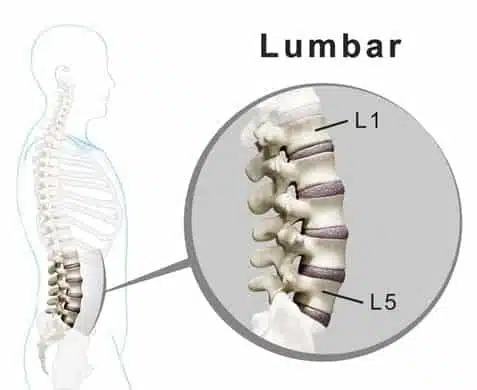
L5-S1 counts as one spinal level: one disc, with two small facet joints behind it that guide movement. Ligaments hold the segment together, and the L5 and S1 nerve roots pass close by. These structures share a small space. So one MRI report can list a disc bulge, enlarged facet joints, a thickened ligament and a narrowed nerve opening at the same level.
L5-S1 also works with the sacroiliac joints and the hip muscles whenever a person stands, walks, lifts or turns. That is why the examination looks at the hips and pelvis as well as the disc.
How L5-S1 Differs From L4-L5
L4-L5 is the level just above L5-S1. Both carry a heavy load, and both show up often in lumbar MRI reports, but they are not built the same way.
L4-L5 sits within the moving part of the lower back, while L5-S1 is where the spine joins the sacrum and pelvis. The disc, the facet joints and the nerve openings (foramina) are angled differently at the two levels. A different pair of nerve roots passes each one.
Symptoms alone cannot reliably tell the two levels apart. L4-L5 findings are often linked with the L5 nerve root. L5-S1 findings can involve the S1 root or the exiting L5 root, depending on where the narrowing sits. So strength, reflexes, feeling, the path of the symptoms and the exact MRI location are compared together.
When a report lists both levels, they do not automatically need the same care. One level may be a chance finding that causes no symptoms, both may contribute, or another structure may matter more. The L4-L5 disc guide explains the level above.
L5-S1 Symptoms: Back, Buttock, Leg and Foot Signs
In my experience, an L5-S1 disc problem often begins as an ache or a cramp-like feeling low in the back or in one buttock. As a nerve root becomes more irritated, the pain can turn into numbness and tingling, then weakness. Leg symptoms usually affect one leg, sometimes both. A tear in the disc wall can make coughing, or turning in bed at night, sharply painful.
People with an L5-S1 disc problem describe it in different ways:
- pressure, stiffness or an ache low in the back, sometimes shifting toward one hip
- pain in the buttock or down the back of the thigh
- tingling, numbness, burning or heaviness in the calf, ankle or foot
- a weaker push-off when walking, or trouble rising onto the toes on one side
The pattern can change from day to day with sitting, standing, walking, bending, lifting and sleep position, and other conditions can look similar. A symptom map is a starting point for questions, not a diagnosis.
Related reading: lower-back guide, sciatic nerve pathway guide, and leg symptom guide.
S1 and L5 Nerve Root Compression: Where the Symptoms Go
Two nerve roots pass the L5-S1 disc. The S1 root crosses the back of the disc on its way down. The L5 root leaves the spine through the opening at its side. When disc material or a bony change presses on either root, an MRI report may call it nerve root compression.
A 2019 review shows an example: a large L5-S1 disc, just off center, pressing on the S1 nerve root (Berry 2019). Disc material out in the side opening can press on the L5 root instead.
Each root has its own area of skin and its own muscles, and the examination uses these maps to find which one is involved:
| Sign | S1 root | L5 root |
|---|---|---|
| Where pain, tingling or numbness can be felt | The back of the thigh and calf, down to the heel and the outer edge of the foot | The outer side of the shin to the top of the foot |
| Movement that can weaken | Pointing the foot down and rising onto the toes, using the calf muscles | Lifting the big toe and turning the foot outward |
Some signs can come from more than one level. Lifting the front of the foot relies mainly on L4, and lifting the big toe on L5. So the examination tests these movements rather than guessing from where the symptoms are felt.
Why Symptoms Can Travel Into the Leg
The L5 and S1 nerve roots run from the spine through the buttock and down the leg. A herniated disc, enlarged facet joints or a thickened ligament can crowd or irritate a root where it leaves the spine. The symptoms are then felt along the path of the nerve, far from the spine. That is why the calf or foot can hurt more than the lower back.
See the nerve symptom guide and pinched nerve guide for broader explanations.
Why Sitting, Standing and Walking Can Feel Different
Position changes the load on the disc, the facet joints and the nerve spaces. A central disc herniation tends to start more abruptly and to hurt more with sitting. Narrowing of the canal tends to hurt more with long standing and to ease with sitting (Lee 2015). The section on facet and ligament changes explains that second pattern.
The useful observation is the pattern: what changes with sitting, standing, walking, bending, coughing, lifting or rest. Short movement breaks, supported sitting and careful lifting limit the load that builds up over a day. The daily habits section gives the intervals used at CSC.
Why the L5-S1 Disc Bulges or Herniates: Causes and Risk Factors
In my experience, most damage to the L5-S1 disc follows an injury, sudden or repeated, or harmful strain repeated over time. A heavy lift, a fall or a sports injury can tear the disc’s tough outer ring at once. Repeated strain works more slowly, through months or years of long sitting, lifting, bending and strenuous work.
Poor habits supply most of that strain. Sitting brings the weight of the upper body down onto the lower discs, and leaning forward adds to it. Bending and twisting at the waist load the disc further, above all when the two come together.
Age is one factor, but not the only one: genetics, lifestyle, posture and past injuries count too. I have seen disc desiccation, a dried-out disc, in a girl as young as seven, and healthy discs in people in their 80s.
L5-S1 sits where the moving lower back meets the pelvis, so it takes part in every lift, bend and turn. Most forward bending happens at L4-L5, which is why I see disc problems there most often, with L5-S1 second. The daily habits section shows how to limit the strain.
Pregnancy and L5-S1
During pregnancy the growing abdomen shifts the body’s center of mass, and the pelvis adapts. The load on L5-S1 changes, so an earlier lower-back or disc problem can become more noticeable. Back and pelvic pain in pregnancy has many possible causes, so it is assessed in person rather than guessed from a web page.
Care stays within scope and is coordinated with the maternity team when needed. These need obstetric or medical assessment first:
- abdominal symptoms, bleeding or fever
- urinary concerns
- sudden swelling or a severe headache
- reduced baby movements
- any concern the obstetric team raises
Related reading: pregnancy-related lower-back guide, pregnancy and back guide, and pelvic symptom guide.
Common MRI Terms at L5-S1
MRI reports describe a disc with words such as bulge, protrusion, prolapse, herniation, extrusion and sequestration. To me these are stages of one process, and slipped disc is the everyday name for all of them. The stages are set out below in my order, and the table explains the other words a report often uses. Whether a finding explains the symptoms is decided at the examination.
| MRI term | Plain-language meaning |
|---|---|
| Disc degeneration or desiccation | The disc has lost some of its water, looks darker on the scan or has lost height. Age, genetics, past injuries, posture and daily habits all play a part, and it may or may not cause symptoms. |
| Bulge, protrusion, prolapse, herniation, extrusion, sequestration | Stages of one process, from the mildest to the most serious, each defined in the stages below. |
| Annular fissure or tear | A split in the tough outer wall of the disc, often seen as a bright spot on the scan. It can be present with or without symptoms and does not decide the care by itself. |
| Foraminal narrowing | Less room in the side opening where a nerve root leaves the spine. The amount, the exact place and the nerve tests decide whether it matters. |
| Central or lateral-recess narrowing | Less room in the middle of the spinal canal (central) or in the side channel next to it (lateral recess). The disc, the facet joints and a ligament can all play a part. |
Protrusion, Herniation and Extrusion: Stages of Disc Damage
Each stage is a progression of the one before. A bulge follows a damaged disc, a herniation follows a bulge, and an extrusion follows a herniation. In my order, with what each means at L5-S1:
- Damaged or degenerated disc: torn, weakened fibers in the outer ring stop holding the soft center firmly in the middle.
- Disc bulge: the outer fibers swell more than usual while the center stays put. A true bulge leaves the thecal sac and the S1 and L5 roots alone.
- Protrusion: the center migrates outward, toward the canal or a side opening, without breaking through the outer fibers. It is still contained.
- Prolapse: the disc is barely contained. Only a few outer fibers keep the center, the nucleus pulposus, from rupturing into the canal.
- Herniation, also called a rupture: the last intact fibers tear and the center breaks through the outer wall, the annulus fibrosus. At L5-S1 it can now touch the S1 root, or the L5 root in the side opening.
- Extrusion: the center is pushed from the middle of the disc out to its edge. A large herniation like this needs prompt assessment, which does not automatically mean surgery and can mean conservative care.
- Fragmentation and sequestration: in my view the last and most dangerous stage. Part of the center breaks off and floats free in the spinal canal.
Each drawing below shows a lumbar disc from above. The front of the body is at the top; the nerve sac and roots sit behind the disc.


L5-S1 Disc Bulge: What It Means
An L5-S1 disc bulge is the mildest stage: a contained slipped disc whose outer fibers have swollen more than usual.
Think of a jelly donut pressed gently from above. The filling stays inside, but the sides push out a little.
The first question I ask is whether the disc is pressing on anything yet. A true bulge has not squeezed or moved a nerve root or the thecal sac, the sleeve around the nerve roots. I am firm on this: a disc bulge, as an MRI shows it, does not compress nerves.
By my definition, a disc that squeezes the thecal sac or the S1 root is a prolapse or a herniation, whatever the report calls it. The word bulge is often used loosely, even in some scan reports, so the examination decides what a finding means.
Bulges are also common in people with no back pain. A 1994 study scanned the lower backs of 98 adults who had none. More than half (52 percent) showed a bulge at one level or more, and 27 percent a protrusion (Jensen 1994). I rate seriousness from 0 to 10. A true bulge rates 1 or 2, and a disc that reaches the S1 or L5 root rates 3 to 5.
When leg symptoms come with a bulge on the report, I explain it as a disc bulge in transition. The MRI is taken lying down, often after a night’s rest has let the disc draw in fluid. Lying flat, its fibers may not reach the nerves, so the report says bulge.
Upright, after hours of sitting or driving, the weight of the upper body can push the fibers further out. The disc then behaves as a prolapse, a protrusion, a herniation or even a focal extrusion, and can press on the S1 root. In that state it is a serious condition.
When the findings match the symptoms, care may include gentle chiropractic care without twisting, spinal decompression, focused physiotherapy and rehab exercise. The plan changes as the back and leg respond.
Why MRI Findings and Symptoms May Not Match
Disc wear, bulges and small tears in the disc wall are common on the scans of people with no symptoms. As I often say, the most common symptom of disc damage is none at all. So the images are read together with the history, the nerve examination, movement and the exact location of each change.
The reverse is also possible: a person can have strong symptoms with a scan that looks mild. A small finding in a nerve opening may matter more than a larger one elsewhere that does not match the symptoms. Position matters too. Most lumbar MRI scans are taken lying down, while symptoms may show more when standing or walking.
Questions that connect a scan to the person include:
- Does the side of the finding match the side of the symptoms?
- Does the level match the nerve signs?
- Are strength, reflexes, feeling or walking measurably changed?
- Could another spinal or non-spinal problem explain the symptoms?
- Would a new scan change the decision?
The table sums up how an L5-S1 finding is read:
| An L5-S1 finding may mean | It does not automatically mean |
|---|---|
| A structural change is visible at L5-S1. | The finding is automatically causing every symptom. |
| The space around a nerve may be reduced. | The amount of narrowing predicts how strong the symptoms are. |
| Several structures may be involved together. | One label or one method explains the whole picture. |
| A focused assessment may be useful. | A website or MRI report can diagnose the reader. |
| Medical or specialist input may be appropriate in some situations. | Surgery is automatically required. |
| The plan reflects the history, the nerve findings, daily function and the person’s preferences. | One MRI term determines the same plan for everyone. |
How an MRI Report Is Read in Context
An MRI report is the radiologist’s summary of the images. It usually covers alignment, disc height and signal, bulges or herniations, the central canal, the nerve openings, the facet joints and the ligaments.
Words such as mild, moderate and severe describe how a change looks, not how much it will affect daily life. Different radiologists may word the same scan slightly differently, so the report is read together with the images and the examination.
Other Findings Often Reported at L5-S1
An L5-S1 MRI report often lists more than one finding. Endplate (Modic) changes, a forward slip of L5, enlarged facet joints, a thickened ligamentum flavum and general wear can sit alongside a disc change. The sacroiliac joint just below the segment can cause overlapping symptoms too. Each one is read the same way as a disc finding: by its type, its exact location and whether it matches the examination.
Modic Type 1 and 2 Changes
Modic changes are changes in the bone marrow next to a disc, seen on MRI. Type 1 points to more active change at the endplate, the thin layer between the bone and the disc. Type 2 shows marrow that has partly turned to fat, a calmer stage.
Modic changes describe what the scan shows; they are not a diagnosis on their own. A review of 31 studies found their link with low back pain inconsistent (Herlin 2018). So a Modic change is not proof that the endplate is the only source of a person’s symptoms.
At L5-S1, Modic changes can appear with a worn or herniated disc, lost disc height or an uneven endplate. The type, the location and the findings around it are noted, then compared with the history and the examination. On its own, a Modic change does not show instability, infection or a fixed outlook.
A medical assessment is added when something points to more than disc wear. That can be the scan itself, fever or weight loss, blood tests or the way the problem behaves over time.
Read the dedicated Modic Type 1 guide and Modic Type 2 guide for fuller explanations.
Spondylolisthesis at L5-S1
Spondylolisthesis means one vertebra has slipped forward on the one below. At L5-S1, the slip may come from wear, a crack in the pars or another structural problem. The pars is a small bridge of bone at the back of the vertebra.
Reports grade how far the vertebra has slipped, but the grade alone does not decide the symptoms or the next steps. Some low-grade slips stay stable, while others come with narrowed nerve openings, disc changes or nerve symptoms.
A useful assessment considers whether the shift is stable and whether symptoms change with position. It also checks for measurable weakness or changes in feeling, and for crowded nerves on the images. The aim of care is not to push L5 back into place. The priority is to understand stability, nerve function and daily function, and to choose care from there.
In my experience, a slipped vertebra turns up far more often at L5-S1 and L4-L5 than higher in the lumbar spine. Still, a slip of any kind can occur at every lumbar level. It usually comes together with disc and joint wear, enlarged facet joints, a thickened ligamentum flavum, and narrowing of the spinal canal and foramina.
When an X-ray shows a slip in either direction, forward (anterolisthesis) or backward (retrolisthesis), CSC may add three side-view X-rays of the lower back. They are taken in a neutral position, bending forward and leaning back, to see whether L5 shifts on the sacrum as the spine moves.
An MRI may follow to show what the slip is doing to the discs, joints, ligaments, nerve openings, canal and thecal sac. A specialist opinion is sought when the findings call for one.
A dedicated explanation is available on the spondylolisthesis guide.
L5-S1 Degenerative Disc Disease: What the Label Means
Degenerative disc disease is a common label for wear in a disc. An L5-S1 report may call the same change disc degeneration or desiccation. The disc has lost water, looks darker on the scan or has lost height.
In my experience, most wear-related changes at L5-S1 follow an injury, sudden or repeated, or repeated harmful strain. Age plays a part but is not the only factor, so degenerative changes can begin far earlier than most people expect.
So the label describes how the disc looks, not whether it causes symptoms. A degenerated disc matters when it narrows the space for a nerve or matches the symptoms, and the examination, not the scan alone, shows that. The degenerative disc disease guide covers the condition across the spine.
Disc Space Narrowing at L5-S1: Why a Mild Finding Can Matter
Many L5-S1 reports describe “mild disc space narrowing” or “loss of disc height.” Mild describes how the disc looks on the scan, not how much the change matters.
As the disc thins, three things can follow:
- more of the load at L5-S1 passes to the facet joints behind it, which can develop arthritis and enlarge
- the ligamentum flavum slackens and can buckle into the canal, narrowing it
- the weakened disc becomes more prone to tears, bulges and protrusions
In my experience, a narrowed disc can affect the L5 or S1 nerve roots before it causes much back pain. The first signs may be numbness or weakness in the leg, or difficulty standing or walking for long. So a mild narrowing is read together with the nerve examination, not set aside because of the word. Treatment for disc degeneration and disc space narrowing at L5-S1 follows that examination, as the treatment section explains.
Facet Hypertrophy, Ligamentum Flavum Thickening and Spinal Stenosis
Facet joints sit behind the disc and guide spinal movement. Facet hypertrophy means these joints have grown larger, often alongside disc wear. Enlarged facets can take up room in the side channel of the canal or in a nerve opening. How much that matters depends on where the change sits, how large it is and whether the nerve tests match.
The ligamentum flavum is an elastic ligament along the back of the spinal canal. Repeated load and scarring over the years can thicken it, and a thinning disc can make it buckle inward. Either change can narrow the canal or its side channels, above all when the facets have enlarged too.
Narrowing of the canal or a nerve opening is called spinal stenosis. It has a typical pattern, called neurogenic claudication. Leg symptoms come on with long standing or leaning back, and they ease with sitting, bending forward or lying down (Lee 2015). The pattern alone cannot confirm stenosis, so the scan is compared with the nerve tests, walking tolerance and other possible causes.
Related guides: facet hypertrophy, ligamentum flavum hypertrophy, and spinal stenosis.
Sacroiliac Joint Overlap
The sacroiliac (SI) joints sit just below the L5-S1 segment and pass load between the spine and the pelvis. A person may feel symptoms near one side of the lower back, buttock, or upper thigh when the SI region is involved. Turning in bed, climbing stairs, standing on one leg, or moving from sitting to standing can change the pattern. These features can overlap with disc, facet, hip, and nerve-related symptoms.
An online symptom list cannot tell an SI problem apart from an L5-S1 problem. The examination compares:
- movement of the lower back and the hip
- the nerve tests
- tasks that load the pelvis
- a group of SI tests that try to bring on the pain
No single test decides it. Both problems can also be present at once.
See the sacroiliac joint guide for a focused explanation.
What Is Lumbar Spondylosis?
Spondylosis means wear-related change in the spine. The term has many definitions, and used broadly it covers the discs, the vertebrae and the joints between them (Middleton 2009). At L5-S1 the word may mean a worn disc, bony spurs at the edges of the vertebrae (osteophytes) or arthritic facet joints. Because the segment carries a heavy load, these often appear together.
“Slipped disc” is an everyday phrase, not an MRI diagnosis. It can mean a bulge, a protrusion, an extrusion or another disc change. The useful step is to name each structure on the report and check whether it matches the examination.
A disc can lose height without pressing on a nerve. A facet can enlarge without producing leg symptoms. A bony change can be present without being the main reason a person struggles day to day.
The spondylosis guide explains the term across the spine.
Teenagers can have low back or leg pain too. Their parents can read the guide to chiropractic care for children. It explains how the L5-S1 region and the rest of a growing spine are assessed.
When a Doctor or Hospital Comes First
Most L5-S1 disc problems can be assessed calmly at a planned visit. Leg weakness that keeps getting worse should not be ignored. It calls for a careful assessment by a competent provider, and conservative care can continue only while the weakness itself is improving.
For back or leg pain associated with an L5-S1 herniated disc, urgent medical attention, a thorough assessment at a hospital emergency department straight away, within hours, is needed if numbness develops or worsens around the genitals, between the genitals and anus, or around the anus (Gardner 2011). New difficulty passing urine needs the same urgent assessment; loss of bladder or bowel control does not have to occur first, and if it does occur, it is urgent too. These symptoms have several possible causes. Some may call for an invasive procedure, but the assessment determines what care is appropriate, rather than assuming surgery is necessary.
A few other signs call for a doctor before spinal care:
- a severe injury, such as a bad fall or a road accident
- back pain with a fever, or with feeling hot, shivery and generally unwell
- weight loss without trying, or back pain that is worse at night
When one of these is found, a medical referral comes first.
What the First Assessment Covers
The assessment starts with a long conversation about the symptoms, earlier episodes, other health conditions, medication, work, exercise and sleep. This history shows whether care at CSC suits the problem or whether a referral should come first.
From arrival to leaving, a first assessment takes 30 minutes to two hours, depending on how complex the problem is. I do not rush it, because a problem that seems mild sometimes needs other areas checked too. It helps to write the history down beforehand, with everything tried so far and the questions to ask. Any MRI or X-ray is worth bringing on CD or film, not only as a report.
Low back pain is among the problems seen most often at CSC. Pain that starts at L5-S1 often travels into the buttock and down the back or outer side of the thigh. Many structures can produce that picture, so the questions work through the possibilities:
- strained muscles or ligaments
- facet joints that press together (imbrication) or have enlarged
- a worn, narrowed disc (disc degeneration)
- disc material that bulges, protrudes, prolapses, herniates, extrudes or breaks off
- a segment that moves more than it should
- a vertebra that has slipped forward or backward, with or without a pars defect (spondylolysis)
- a thickened ligamentum flavum
- narrowing of the canal or of a nerve opening (foramen)
- pressure on the L5 root as it exits, or on the S1 root as it passes the disc
The physical examination looks at walking and movement, lumbar and hip motion, and the nerves: strength, reflexes and feeling, with tests chosen for the person. No single test settles the source. The examiner looks for findings that fit together, and for any that change the urgency or call for referral first.
Nerve findings are read as a group, and weakness that is new or getting worse matters most. The same tests are repeated at later visits, so any change in nerve function is seen early.
X-Ray or MRI: How the Choice Is Made
Imaging is not routine at CSC. It is added when its result will change the plan, and one study is usually enough; an X-ray and an MRI together are the exception. What the examination points to decides which is used:
| What the examination points to | Imaging used at CSC |
|---|---|
| Alignment, the soft tissues or a facet joint problem (facet joint syndrome) | An X-ray may be enough |
| A disc problem, from a bulge, protrusion, prolapse or herniation to an extrusion or fragment | An MRI, so that a disc diagnosis is confirmed on imaging rather than guessed |
| A step felt between the bony points along the spine (the spinous processes), suggesting a slipped vertebra such as a grade 1 or 2 spondylolisthesis with a pars defect | Both an X-ray and an MRI |
Existing MRI or X-ray images are reviewed together with the written report. The reason for any new scan or referral is explained before the person goes ahead. An MRI shows the disc, the nerves and the space around them; a plain X-ray shows the bones and how they line up.
What Is the Best Treatment for an L5-S1 Disc Bulge?
No single treatment is best for every L5-S1 disc bulge. The choice depends on what the examination finds, how irritated the nerve is and how long the symptoms have lasted.
In my view, any care starts with an accurate diagnosis and a proper plan. It then needs careful delivery, with as few flaws as possible, and changes to posture, habits and daily activity. An L5-S1 plan can be checked against these points:
- An examination first: calf strength, the ankle reflex and feeling in the outer foot, with any MRI read against them (see the first assessment).
- Realistic goals: a damaged disc does not return to its original state. The plan aims at how well the back and leg cope with daily life (see healing without surgery).
- Methods that spare an irritable disc: gentle, non-twisting methods, with no bending, twisting or forceful manipulation while the disc is sensitive (see the treatment section).
- A check of the neighbors: the sacroiliac joint and the hip can cause similar pain, so both are examined (see sacroiliac joint overlap).
- Sitting and daily load: long sitting tends to worsen disc pain, so the plan includes movement breaks and supported sitting (see daily habits).
- Measured progress: the first visit’s tests are repeated at each review, and the plan changes if they stall (see recovery time).
- Surgery only in set situations: an international spine committee recommends it when conservative treatment has not worked or nerve function keeps getting worse (Costa 2024). The section on when surgery is considered gives the full list.
Before choosing, it helps to call or WhatsApp the centers being considered. Describe the L5-S1 findings and ask how each would examine them.
Treating an L5-S1 Herniated Disc Without Surgery
Care for an L5-S1 disc problem is chosen after the assessment, not from the MRI label alone. A bulge, a tear in the disc wall, a Modic change or a thickened ligament does not by itself decide which method suits.
The plan weighs nerve function, how easily symptoms flare, health history and how earlier care went. Work and family demands count too, along with the person’s goals and preferences.
Chiropractic and physiotherapy remain separate professional services, even when they are coordinated. A registered chiropractic practitioner is responsible for the chiropractic care, and a registered physiotherapist for the physiotherapy. I recommend the two together rather than one or the other, and a referral elsewhere comes first when the findings call for it.
How CSC Cares for a Lumbar Disc Problem
The care comes from a combined team of chiropractors and physiotherapists, and each method is chosen for the tissue that needs it. The range includes:
- spinal decompression therapy
- flexion-distraction (the Cox technique)
- ultrasound, electrotherapy and shockwave
- high-intensity laser therapy
- cold therapy with the QMD cryothermal device (the device’s heat setting stays off any spine with a disc problem)
- trigger point and myofascial therapy
- stretching, strengthening, balance and neurological exercises, each at the right stage
- devices for pain management
Some things are deliberately left out while an L5-S1 disc is irritable. Bending and twisting are avoided. So is any rotational or forceful physiotherapy, manual therapy or chiropractic manipulation, since twisting can aggravate a disc problem. I am not a fan of rotary chiropractic manipulation, and I use it only when I am certain there is no disc damage. Cracking and twisting should not be done on a spine with a damaged disc, however experienced the hands.
Every session stays within what the patient can comfortably tolerate. Patients are asked to speak up at once if anything feels uncomfortable, because discomfort during care can turn into more inflammation and pain later. My motto for this is “No pain, more gain.”
Stretching and exercise wait until the back is stable and ready. For most people that falls between the 5th and 7th session, and for some it comes as early as the 3rd. The first exercises depend on how severe the problem is and usually draw on:
- Kegel exercises when the pelvic floor is weak
- wall-assisted squats
- the cat and camel movement
- core canister exercises for the deep trunk muscles
- strengthening for the glutes, hamstrings, calves and quadriceps
An exercise that brings on more nerve symptoms or weakness is changed rather than pushed through. Progress follows the person’s response, not a fixed timeline.
For the gym, the advice is to stay away at first and to leave free weights alone for 2 to 6 months. Once the back has stabilized, some machine work can return:
- hip adductor and abductor machines
- leg curls and leg extensions
- light rowing and lat pulldowns
- for some people, the chest press
Deadlifts, leg presses and free weights stay out. Treadmill walking is kept flat, with no incline, because walking uphill puts more load through the lumbar discs.
Age changes the plan as well. When an older adult’s L5-S1 problem involves stenosis or arthritis, care moves more gently:
- more emphasis on adjusting daily habits
- stretching introduced later
- extra stability work on the rocker board, the wobble board and a gym ball, with Kegel exercises
- lighter and shorter decompression sessions
- gentler manual and myofascial work
A younger adult with an L5-S1 bulge more often follows the usual order: calming the irritated tissue first and building strength afterward.
When Surgery Is Considered
Surgery has its place, but with a herniated or bulging disc it is the last option, once the conservative route has been exhausted. In my practice, an operation is raised in three situations:
- a conservative plan that is not working
- a severe case, such as a sequestered disc or an infection
- the urgent signs that need a hospital emergency department
An international spine committee recommends surgery in similar situations (Costa 2024):
- conservative treatment that has not worked
- cauda equina syndrome, the rare problem behind those urgent signs
- nerve function that keeps getting worse, including severe weakness
The same review does not recommend spinal fusion as a routine treatment for a first lumbar disc herniation. It may still be considered in some cases, such as instability.
The decision is made individually, weighing the benefits, the limits, the alternatives and the person’s own preferences.
Learn about physiotherapy services and the main CSC services overview.
L5-S1 Disc Bulge Recovery Time: How Long It Takes to Heal
There is no single timeline, because each person heals differently. The pace depends on how large the herniation is and whether the nerve is weak or only irritated. How long symptoms have lasted, and daily habits such as sitting and lifting, matter too. Leg pain, numbness and strength can each settle at a different speed.
The question I hear most often is whether a full recovery is possible. A damaged disc does not return to its original state, so I judge recovery by how the back and leg work, not by the scan. In my experience, an L5-S1 problem left untreated for a long time more often leaves lingering symptoms or flare-ups, even after a good recovery.
Progress is counted in weeks, not days. At each review, CSC repeats the tests from the first visit. The person rates their pain on a visual analog scale (VAS) and describes how daily life is going.
Without improvement by the fifth session, a reassessment is due. If there is still none by the 10th, continuing or stopping is a decision made together with the chiropractor. Surgery stays an option if symptoms keep getting worse or there is no improvement after a fair trial.
Symptoms and function can improve before a repeat scan shows any change, and a finding can stay the same size while the person recovers. So progress is judged by walking distance, sitting tolerance, sleep, foot control and daily activities. A repeat scan is considered when its result would change the plan. When the retested findings stop changing, the active plan ends at MMI, as the section on healing without surgery explains.
Daily Habits That Ease the Load: Sitting, Lifting, Sleep and Travel
This is the daily advice given at CSC while an L5-S1 disc is irritable. A session lasts from 30 minutes to two hours, and the other 22 hours of the day decide how the disc is loaded. An assessment comes first when symptoms are severe, getting worse or affecting the nerves.
- Sitting: with acute back pain, a break every 15 minutes to stand and walk for 10 to 20 seconds. Without back problems, a break every 30 to 45 minutes. No slouching over a desktop or laptop.
- Chair: one that supports a comfortable position with the feet resting securely; even a good posture is broken up at each break.
- Bending, twisting and lifting: kept to zero while the disc is irritable. Anything that cannot wait weighs under 2 kg and is held close, with the hips and knees bending.
- Stretching: no aggressive stretch that chases a nerve feeling down the leg, and nothing that bends or twists the spine deeply.
- Yoga: it must be avoided at every stage of an L5-S1 disc problem, not only while the disc is irritable.
- Activity: increased gradually, since a sudden jump in lifting, running, gardening, travel or gym work can exceed what the back can take.
- Sport: resumed after recovery. Anything that hurts at the time, or brings on more pain within 72 hours, is left out, since inflammation can take that long to show. Lap swimming waits as well; walking in water is the gentler choice.
- Travel: long road trips postponed where possible. A passenger rides in the reclined front seat, and a driver stops every 45 to 60 minutes to walk and stretch the calves.
- Sleep: at least six hours a night, and seven to eight hours unbroken where possible. Among positions that do not hurt, the right side comes first, the left side second and the back third, with a bolster under the knees.
- Ice: 2 to 3 times a day for 10 to 15 minutes, at least 2 hours apart. The pack is wrapped in a cloth, with a timer running. Hot packs are left out, since heat can add to inflammation.
Sciatica vs Slipped Disc: What the Video Explains
To me, sciatica is not a diagnosis. It describes a symptom, an inflamed sciatic nerve, which is formed from several lower spinal nerve roots. It is a nerve problem, not a muscle problem, and in every case I have seen there was some disc involvement, from mild to moderate. The assessment finds which level and which structures are involved.
Read the main sciatica guide and watch the educational video on sciatica and disc-related leg symptoms.
For the chapter list and the transcript, open the Sciatica vs Slipped Disc video page.
Topics in the video:
- 00:00 why sciatica is a symptom, not a diagnosis
- 00:27 how lower spinal nerve roots form the sciatic nerve
- 02:36 why I hold that every case of sciatica involves the disc
- 03:20 how the muscles and fascia react to an irritated nerve
- 05:03 breaks from sitting, walking and side sleeping
- 08:10 stretching only when ready, and nothing too aggressive
- 10:05 why cracking and twisting should not be done on a damaged disc
- 10:57 surgery as the last option
- 11:30 condition-based care and the other 22 hours of the day
Fees for a first visit and for ongoing care are on the chiropractic price in Kuala Lumpur and physiotherapy fees in Kuala Lumpur pages. The fee for a plan is explained and agreed before care begins.
Disc Bulge and Herniation FAQ
Can an L5-S1 disc cause bladder or bowel problems?
Rarely, but it is one of the urgent signs to know. The rare condition behind these signs, cauda equina syndrome, most often follows a large central disc herniation (Gardner 2011). New numbness around the genitals or anus, new difficulty passing urine, or loss of bladder or bowel control needs urgent medical attention at a hospital emergency department straight away, within hours. Either sign is enough on its own, and bladder or bowel control does not have to be lost first.
Can an L5-S1 disc cause hip or knee pain?
It can feel that way: pain from L5-S1 often spreads into the buttock and the back or side of the thigh, close to the hip. In my experience, it can reach the front of the thigh too. If a nerve root is involved, symptoms follow it past the knee to the calf, shin or foot (Berry 2019). The hip joint and the sacroiliac joint can cause similar pain, so the examination checks both to find the source.
Which exercises should be avoided with an L5-S1 disc bulge?
While the disc is irritable, avoid exercises that bend or twist the spine, and any stretch that chases a nerve feeling down the leg. At the gym, deadlifts and leg presses stay out, free weights are left alone for 2 to 6 months, and treadmill walking stays flat. Yoga is avoided at every stage of an L5-S1 disc problem. Exercise begins once the back is stable, for most people between the 5th and 7th session. Any exercise that brings on more nerve symptoms is changed.
Is walking good for an L5-S1 disc bulge?
Yes, while it stays comfortable. Shorter, more frequent walks on flat ground are often easier than one long walk while the disc is sensitive. Walking also breaks up sitting: with acute back pain, stand up to walk every 15 minutes, and without back problems every 30 to 45 minutes. If leg pain builds with walking and eases with sitting, the examination checks for narrowing around the nerves.
How should I sleep with an L5-S1 disc bulge?
Aim for at least six hours a night, and seven to eight hours of unbroken sleep where possible. A short night is not made up with an afternoon nap. Pressure inside the disc is lowest while lying down, and that is when it takes in fluid and nutrients. Among positions that do not hurt, I suggest the right side first, the left second and the back third, with a bolster under the knees. On the side, keep the spine in one line, with a pillow for the neck and a bolster between the knees. Use the left side if the right side brings on heartburn.
Is sciatica a nerve or muscle problem?
A nerve problem. To me, sciatica describes an inflamed sciatic nerve, and every case I have seen came with some disc involvement, from mild to moderate. The muscles and fascia of the back, buttock, hamstring and calf share that nerve supply, so they can tighten and develop trigger points as well. That is why the assessment looks at the disc, the nerve and the muscles together, and why care addresses all three.
Is chiropractic safe with a herniated disc?
In my view, chiropractic care is safe when a competent, registered chiropractor performs it and it is condition-based. Not all chiropractors are equal, and problems come from the practitioner’s choices. For an irritable L5-S1 disc, I use gentle methods without twisting, as the treatment section explains. Bending, twisting and forceful or rotational manipulation are left out.
How to Read This Guide
Everything on this page is general information about the L5-S1 disc. Only an examination can show what is causing one person’s symptoms, what their scan means and which care suits them.
References
- Berry JA, Elia C, Saini HS, Miulli DE. A Review of Lumbar Radiculopathy, Diagnosis, and Treatment. Cureus. 2019;11(10):e5934.
- Costa F, Oertel J, Zileli M, et al. Role of surgery in primary lumbar disk herniation: WFNS spine committee recommendations. World Neurosurg X. 2024;22:100276.
- Gardner A, Gardner E, Morley T. Cauda equina syndrome: a review of the current clinical and medico-legal position. Eur Spine J. 2011;20(5):690-697.
- Herlin C, Kjaer P, Espeland A, et al. Modic changes-Their associations with low back pain and activity limitation: A systematic literature review and meta-analysis. PLoS One. 2018;13(8):e0200677.
- Jensen MC, Brant-Zawadzki MN, Obuchowski N, et al. Magnetic resonance imaging of the lumbar spine in people without back pain. N Engl J Med. 1994;331(2):69-73.
- Lee SY, Kim TH, Oh JK, Lee SJ, Park MS. Lumbar Stenosis: A Recent Update by Review of Literature. Asian Spine J. 2015;9(5):818-828.
- Middleton K, Fish DE. Lumbar spondylosis: clinical presentation and treatment approaches. Curr Rev Musculoskelet Med. 2009;2(2):94-104.
These references describe lumbar disc problems, their imaging and their care in general. None of them evaluates care at Chiropractic Specialty Center.
The Author of L5-S1 Disc Bulge & Herniation: Symptoms, Causes and Care
L5-S1 Disc Bulge & Herniation: Symptoms, Causes and Care was written by Yama Zafer, D.C., who has 30+ years in chiropractic and physiotherapy and founded Chiropractic Specialty Center in Kuala Lumpur in 2006.
Last Updated: L5-S1 Herniated Disc: Symptoms, Treatment and Recovery Time
L5-S1 Herniated Disc: Symptoms, Treatment and Recovery Time was last reviewed and updated on October 10, 2026.
Please help me! The surgeon said there's no bone, so they can't put a spacer in. I've already had an intercept surgery, and their last result is now just a pain pump. I don't believe it; I'm tired of taking pain pills and being tied to pain clinics; please help me.
I'm 51, and I don't want to be in a wheelchair or a walker in four or five years. I tried the pain clinics with pain pumps.
Being told there is not enough bone to hold a spacer is worth looking into before you accept a pain pump. At 51, bone that cannot hold hardware is unusual. Ask your doctor for a bone density scan and the blood tests that look for causes of bone loss. Then ask your surgeon whether better bone density would change the options. Treatment for low bone density takes many months, so it is worth asking early.
If the procedure you had did not change your pain, it is fair to ask whether the source of the pain has been confirmed. A pain pump is a significant step with ongoing maintenance, and it is reasonable to want that question answered first.
Staying out of a wheelchair or a walker is a goal worth planning around. Your strength and walking are the part most in your hands. Long rest weakens the muscles. Choose activity that does not provoke your pain, such as a recumbent bike, walking in water or short walks spread through the day. Build it up a little at a time. Avoid anything that hurts at the time or brings on more pain within 72 hours.
If your symptoms change, arrange a medical review. Leg weakness that keeps getting worse should not be ignored. It calls for a careful assessment by a competent provider, and conservative care can continue only while the weakness itself is improving. New numbness around the genitals or anus, new difficulty passing urine, or loss of bladder or bowel control needs urgent medical attention at a hospital emergency department straight away, within hours.
Do you have such centre in Africa?
All of our centers are in Malaysia, so there is none in Africa. What I can share is what to look for wherever you go.
Check that the practitioner is registered where you live, and ask how often they see your condition. The first visit should be a thorough examination before any treatment, with strength, feeling and reflexes tested where symptoms travel into a leg. With a disc problem, ask whether the methods avoid twisting and forceful cracking. Ask how progress will be measured and when the plan is reviewed if it is not working. Avoid committing to a long prepaid course before an examination.
Ask the imaging center for your scans as images on CD or film, or through its portal, as well as the written report. Another radiologist or a spine specialist can then give a second reading from the images without you traveling.
Wherever you are treated, two measures show how things are going: how far your symptoms travel, and how far you can walk. Symptoms that retreat toward the back mean the nerve is less irritated, while symptoms spreading further down mean the opposite.
If your symptoms keep getting worse, arrange a medical review. New numbness around the genitals or anus, new difficulty passing urine, or loss of bladder or bowel control needs urgent medical attention at a hospital emergency department straight away, within hours. Fever or unexplained weight loss with back pain also calls for a medical review.
Sir, I have L3-L4, L4-L5, and L5-S1 problems. Can these be treated at your chiropractic and physiotherapy centre?
Regards,
Fayaz
Findings at L3-L4, L4-L5 and L5-S1 together are common on lumbar reports. The report alone cannot show which of them is causing your symptoms; the examination decides that.
Where your symptoms travel points to the level. L4, from L3-L4: the front and inner thigh toward the inner shin, with weakness straightening the knee. L5, usually from L4-L5: the outer calf to the top of the foot, with weakness lifting the foot or the big toe. S1, usually from L5-S1: the back of the calf to the outer foot, with weakness rising onto the toes.
If both legs feel heavy after walking a certain distance and ease when you sit or lean forward, the canal may be narrowed. Forward-leaning positions, such as walking with a trolley, then let you stay active.
At CSC every case begins with a thorough assessment, and the MRI images on CD or film are worth bringing as well as the report. Care for damaged discs uses gentle methods that do not twist the spine, together with physiotherapy. Exercise matched to the findings follows once the back is stable.
Write down where your symptoms reach and whether that is spreading or retreating. Leg weakness that keeps getting worse should not be ignored. It calls for a careful assessment by a competent provider, and conservative care can continue only while the weakness itself is improving. New numbness around the genitals or anus, new difficulty passing urine, or loss of bladder or bowel control needs urgent medical attention at a hospital emergency department straight away, within hours.
Hi, I would like to ask some questions about my situation. I have L5-S1 spondylolisthesis with nerve impingement on the L5 nerve root. What is the likelihood it is CES? I have minimal pain and some back discomfort. Erectile dysfunction and penile numbness, rectal numbness and others.
No comment can say how likely that is; only an examination in person can. New numbness around the genitals or anus, new difficulty passing urine, or loss of bladder or bowel control needs urgent medical attention at a hospital emergency department straight away, within hours. Mild back pain does not make nerve pressure less likely, so describe every symptom you listed to the doctor.
An L5 nerve root usually gives symptoms down the outer calf to the top of the foot. The genital and rectal area is supplied by the lower sacral roots, S2 to S4, which run lower in the canal. So the finding you describe may not account for these symptoms, which is a reason to have them examined rather than judged from the report.
Ask for the MRI images to be reviewed against these symptoms. Ask also about a bladder scan after you pass urine, which measures how much is left behind. If pressure on those nerves is ruled out, the other symptoms still deserve a medical review, since they have other possible causes.