
Modic Type I Endplate Changes: Meaning, Symptoms and Treatment
Is a Modic Type 1 Change Serious? What the Research Found
How serious a Modic Type I change is depends on what comes with it. Over time the change can settle, spread or stay the same, and its link with pain varies between people. Worsening leg weakness, fever with back pain or one of the urgent signs below makes the picture more serious. Worsening weakness needs a careful assessment, and fever with back pain needs a prompt medical check. New or worsening numbness around the genitals or anus, new difficulty passing urine, or any loss of bladder or bowel control needs urgent medical attention at a hospital emergency department straight away, within hours.
Can Modic Type 1 Changes Settle or Reverse?
Many Type 1 changes turn into the calmer Type 2 stage, and some spread instead. Turning into Type 2 is a different marrow change, not a return to normal bone. The 1988 study that sorted these changes into types rescanned six people with Type 1. In five, it had turned into Type 2 within 14 months to three years. Type 2 changes stayed the same in all ten people followed for two to three years (Modic and colleagues, 1988).
A later study rescanned 48 Type 1 changes in 44 people, one to six years apart. Of these, 18 (37.5%) had fully turned into Type 2 and 7 (14.6%) partly. Another 19 (39.6%) had spread, and 4 (8.3%) were unchanged. Pain and disability scores were lower where Type 1 had turned into Type 2. The difference was too small to rule out chance (Mitra and colleagues, 2004).
A Modic change sits beside a damaged disc. In my view, disc damage is a progressive condition, as I explain in my video Why Spinal Discs Get Damaged. In my talk Back Pain to Leg Pain, I add that once a disc is damaged, the damage can progress rapidly or take years.
Back Pain and Type 1 Changes Over Two Years
In a Finnish study, pain and disability tended to fall over two years as the Type 1 area shrank, and to rise as it grew. The study followed 64 people with chronic low back pain and a Type 1 or mixed Type 1/2 change. On average, the Type 1 share of the changed bone fell from 74% to 41%, while the Type 2 share rose from 24% to 56%.
Pain fell in 41 people and rose in 15, and disability fell in 44 (Järvinen and colleagues, 2015).
Do Modic Type 1 Changes Cause Back Pain?
Modic Type I changes can go with back pain, but not in everyone, and the research is mixed. A 2018 review of 31 studies found the link between Modic changes and low back pain inconsistent. Fifteen studies found a link and one found the opposite. Only one study was at low risk of bias, and the authors expect better studies to change the picture (Herlin and colleagues, 2018).
An earlier review found a link in seven of ten studies. In people with low back pain or sciatica, the median share with a Modic change of any type was 43%. In people drawn from outside clinics it was 6%. The review also noted that Modic changes can be present in people free of back pain (Jensen and colleagues, 2008).
From how a damaged disc looks on a scan, I can often make an educated guess at how long the damage has been there. My talk Back Pain to Leg Pain makes the point that, in most instances, it has been there for decades without pain.
In two studies, Type 1 changes were more closely tied to pain than other types. Among 228 working men, Type 1 changes were more likely than other types to go with pain (Kuisma and colleagues, 2007). In a study of 74 people, 27 of the 37 whose changes were dark on T1, as Type 1 changes are, had low back pain. Only 4 of the 37 whose changes were bright on T1, as in Type 2, did (Toyone and colleagues, 1994).
Antibiotics Against a Dummy Pill: The Norwegian Trial
For chronic low back pain with Modic changes, three months of antibiotics gave no meaningful benefit over a dummy pill in a Norwegian trial. A low-grade infection has been suggested as a cause of Modic changes. The trial tested antibiotics in 180 adults with chronic low back pain, an earlier slipped disc and Modic changes at the same level. Of these, 118 had Type 1 changes. Half took the antibiotic amoxicillin three times a day for three months, and half took a dummy pill.
After one year, scores on a 24-point disability scale differed by 1.6 points. The researchers had set 4 points in advance as the smallest meaningful difference. Among the people with Type 1 changes, the difference was 2.3 points, also short of that mark.
Adverse effects were reported by 56% of the antibiotic group and 34% of the dummy-pill group. The researchers concluded against antibiotics for chronic low back pain with Modic changes (Bråten and colleagues, 2019).
Back and MRI Assessment at Two KL Centers
For a Modic Type I change, a first assessment starts with an interview, then checks movement, strength, reflexes and feeling in the legs. The MRI images are read after that, so bring them on CD or film, not only the reports. Either Kuala Lumpur center can arrange the visit:
- Bukit Damansara (KL, main center): Call +603 2093 1000 or WhatsApp +60 17 269 1873 to see the Bukit Damansara / KL center.
- Bandar Sri Damansara (KL): Call +603 6262 5777 or WhatsApp +60 12 455 6939 to see the Bandar Sri Damansara center.
Directions and hours for both centers are on the contact page. The Bukit Damansara center also serves nearby Taman Tun Dr Ismail (TTDI), Bangsar & Bangsar South, Mont Kiara and Sri Hartamas. It also serves Mid Valley & KL Eco City and Solaris Mont Kiara & Publika. The Bandar Sri Damansara center serves nearby Petaling Jaya.
What Modic Type I Endplate Changes Look Like on an MRI

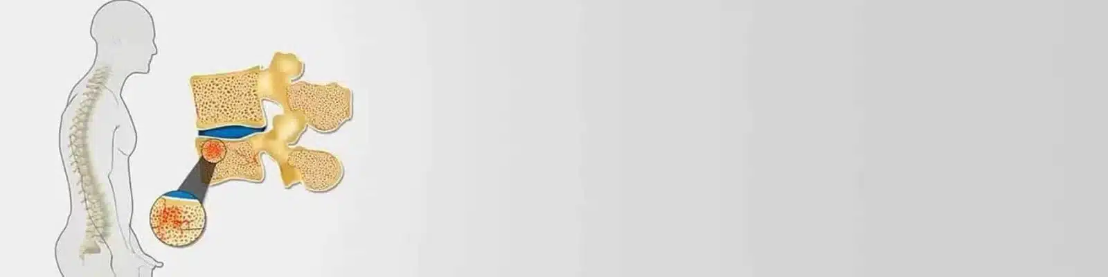
On an MRI, a Modic Type I endplate change shows as marrow beside the endplate that looks dark on T1 and bright on T2. T1 and T2 are the two main kinds of MRI picture, and Modic types are sorted by how that marrow looks on each. This pattern is thought to reflect an inflammatory reaction in the marrow (Herlin and colleagues, 2018).
Inflammation is one of the first things to happen in disc damage, in my view. Inside the disc, it is what breaks the disc down, as I explain in my talk Back Pain to Leg Pain. It also occurs around the spine: in the facet joints, the small joints at the back of each level, and in the deep back muscles. An MRI can show it there.
To me, an MRI is a gold standard for imaging the back, a view I give in my video Is Chiropractic Safe? 13 Questions Answered. It uses no radiation and shows the bones, joints, muscles and ligaments, even the space the nerves take up.
In the 1988 study, which reviewed the scans of 474 people, Type 1 showed up in 4% and Type 2 in 16%. Every change sat at a level with a worn disc (Modic and colleagues, 1988).

Modic Classification: The Three Types Compared
| Type | On T1 | On T2 | What it reflects |
|---|---|---|---|
| Type 1 | Dark | Bright | An active stage: tissue samples show cracks in the endplate and new, blood-rich scar tissue (Modic and colleagues, 1988) |
| Type 2 | Bright | Same or slightly bright | A calmer stage: tissue samples show marrow that has turned to fat (Modic and colleagues, 1988) |
| Type 3 | Dark | Dark | Hardened, thickened bone, called sclerosis (Herlin and colleagues, 2018) |
The guide to Modic Type II changes covers the calmer Type 2 stage in full.

Where Modic Type I Changes Usually Appear
Modic Type 1 changes, like Modic changes in general, appear most often at L4-L5 and L5-S1, the two lowest levels of the lower back. In 228 working men, 80% of all their Modic changes were at L4-L5 or L5-S1. Changes at L5-S1 went with more frequent low back pain and sciatica (Kuisma and colleagues, 2007).
Among 101 people with a Type 1 change at a single level, 37 had it at L4-L5 and 37 at L5-S1. Those with less disc narrowing were younger and more often had the change at L5-S1 (Nguyen and colleagues, 2017).
In my talk Back Pain to Leg Pain, I explain that 95% of bending at the waist happens at L4-L5. On average, that makes it the most common level for disc damage, with L5-S1 second.
The guides to the L4-L5 level and the L5-S1 level explain what else can affect these two discs.
Symptoms of Modic Type 1 Changes
Low back pain is the main symptom that goes with Modic Type 1 changes. In my experience, pain from the lower back often spreads into the buttocks and the thighs.
The pain can follow a recognizable pattern. A study compared 120 people with chronic low back pain, half of them with Type 1 changes. Those with Type 1 changes more often had at least one of three features: night pain, pain worse on waking, or morning stiffness. They were also far more likely to have pain when bending backward. Pain when bending forward was about as common in both groups (Bailly and colleagues, 2014).
Leg Pain and Pinched Nerve Symptoms
When the damaged disc also irritates or presses on a nerve root, pain can travel down the leg, sometimes with numbness, tingling or weakness. This is sciatica, often called a pinched nerve, and the sciatica guide covers it in more depth. My video Sciatica vs Slipped Disc: Video Explanation makes the point that sciatica is a symptom, not a diagnosis. I have yet to see sciatica without some disc involvement.
Leg weakness that keeps getting worse should not be ignored. It calls for a careful assessment by a competent provider, and conservative care can continue only while the weakness itself is improving.
If you have back or leg pain, new symptoms need attention even when an MRI report has already described a Modic Type I change. New or worsening numbness around your genitals, between your genitals and anus, or around your anus needs urgent medical attention: a thorough assessment at a hospital emergency department straight away, within hours. New difficulty passing urine needs the same urgent assessment before bladder or bowel control is lost, and a loss of that control is urgent too. Several causes are possible. Some may call for an invasive procedure, but assessment helps determine the next step and does not automatically mean surgery.
Back pain in a child or teenager calls for an assessment adapted to a growing spine, as the guide to chiropractic care for children explains.
What Causes Modic Type 1 Endplate Changes?
Modic Type 1 changes begin with damage where a disc meets the bone. A disc gets its blood supply from the bone, a point I make in my talk Back Pain to Leg Pain. So the bone has to be healthy for the disc to be healthy. The endplate is the route in: during sleep, when the pressure inside the disc drops, the disc draws in fluid and nutrients through it. In my experience, the damage behind most changes like this comes from an injury, sudden or repeated, or from repeated harmful strain. Age is one factor, never the only one: genetics, posture, daily habits and earlier injuries all contribute.
The endplate is where much of that damage shows. In 54 people with a large Type 1 change, 96% had damage to the endplate beside it. Over the next year, growing or shifting Type 1 changes went with more endplate damage, a thinner disc and changes in the disc on MRI. Discs free of a Type 1 change rarely worsened (Kerttula and colleagues, 2012).
One Sudden Injury or Many Small Strains
A single injury can be a heavy lift, a fall or a sudden twist. Repeated strain builds up over long hours of sitting, slouched at a desk or laptop, and from bending and twisting at the waist. Much of that strain is axial load bearing: the body's weight pressing down through the spine. In my talk Back Pain to Leg Pain, I note that the research I have read names it the most common cause of disc damage. I explain how poor habits damage a disc in my video Why Spinal Discs Get Damaged. Cutting back on these strains is part of every care plan.
After a Slipped Disc
A slipped disc can be followed by new Modic changes at the same level. In 166 people followed for 14 months after severe sciatica, the share with Type 1 changes rose from 9% to 29%. New changes appeared mostly at the level of the earlier disc herniation (Albert and Manniche, 2007).
A disc that has slipped was already degenerated, in my view. As I put it in my talk Back Pain to Leg Pain, no disc bulges or herniates without degeneration first.
A Spinal Segment That Moves Too Much
Extra movement is common at a spinal level with a Type 1 change. A 1994 study of 74 people with these marrow changes took X-rays while each person bent forward and back. Too much movement showed in 70% of those whose changes were dark on T1, the Type 1 look. The same was true of 16% of those with changes bright on T1 (Toyone and colleagues, 1994).
At CSC, when an X-ray shows one vertebra shifted on the next, side-view X-rays taken bending forward and backward may be added to check stability.
Treatment for Modic Type 1 Changes Without Surgery
Treatment for a Modic Type 1 change without surgery is focused conservative care. By conservative care, I always mean focused care, planned from what the MRI and the examination have found. It is aimed at the causes those findings document, takes in the whole lower back and does no harm. Routine back treatment is not what I mean; many forms of it are useless.
At CSC, care for a Modic Type I change keeps to the sequence I have followed over my 30+ years in chiropractic and physiotherapy:
- A detailed history: how the pain began, where it spreads and what changes it.
- An examination of movement, with orthopedic tests, nerve function and muscle strength.
- The MRI, read with those findings, since the diagnosis comes from all three together.
Chiropractors and physiotherapists then plan the care together, matched to the tissues involved. Depending on the findings, it may include:
- Spinal decompression therapy, a gentle, controlled pull along the lower spine.
- Flexion-distraction (the Cox method), a gentle, rhythmic stretch of the lower back on a moving table.
- Chiropractic manipulation that is non-rotatory using the Activator instrument, and other gentle, twist-free chiropractic care, as the guide to non-twisting methods explains.
- Trigger point and myofascial therapy for tight, protective muscles.
- Therapeutic ultrasound, electrotherapy, shockwave therapy and high-intensity laser therapy, chosen for the tissues involved.
- Cold therapy with the QMD cryothermal device. Its heat setting is not used on a spine with a disc or facet joint problem.
- Stretching, strengthening, and balance and nerve-control exercises, once the area has settled.
- Physiotherapy and rehabilitation that build up as the back allows.
At CSC, this care sits within NSD Therapy®, which combines spinal decompression, non-rotatory chiropractic, physiotherapy and exercise. Its decompression part is delivered with the RxDecom® system.
My principle is that recovery has a better chance when what is beneficial for the back increases and what is harmful to it decreases. That takes an accurate diagnosis, a proper plan, execution with no flaws or the fewest possible, and changes to posture, habits and daily activity.
What Is Avoided During Care
With a Modic Type I change, these are left out:
- Bending and twisting at the waist.
- Any rotational or aggressive manipulation, manual therapy or physiotherapy, since twisting can aggravate a disc problem.
- Stretching and exercise in the early, irritable stage, apart from Kegel exercises for the pelvic floor.
- Yoga, which must be avoided, since many of its poses bend and twist the spine.
Care is kept within what you can comfortably tolerate. My video Is Chiropractic Safe? 13 Questions Answered explains why: any discomfort is a sign that care is too aggressive. In that video, I also say that anything beyond non-rotatory methods can create inflammation or worsen disc damage already there. Tell the clinician at once about any discomfort, even when it is slight, since discomfort may also increase inflammation and pain later. My motto has always been “No pain, more gain.”
Daily Habits With a Modic Type 1 Change
Daily habits are part of care for a Modic Type 1 change. In my talk Back Pain to Leg Pain, I put lifestyle change ahead of treatment; with significant pain, the two start together. As I point out in Sciatica vs Slipped Disc: Video Explanation, a visit with me lasts 30 minutes to two hours of a 24-hour day. Posture and habits in the other 22 hours matter too, so I advise these daily changes:
- Stand up from sitting every 15 minutes while the back is acutely painful. As I advise in my video Desk Posture Mistakes, walk for 10 to 20 seconds.
- With acute, intense back pain, limit driving to about 15 minutes at a time, and never go past 20 to 30 minutes. Take each break where it is safe to stop, then walk and stretch the calves.
- Ice the sore area two or three times a day, for 10 to 15 minutes each time and at least two hours apart. Use a timer, and keep a cloth between the pack and the skin.
- Keep hot packs off the lower back. My talk Back Pain to Leg Pain explains why: with a damaged disc or an inflamed area, their surface heat can add to the inflammation.
- Sleep in the least painful position for at least six hours a night. My video Neck and Back Self-Help Tips: Posture, Sleep and Movement explains why sleep matters so much. My talk Back Pain to Leg Pain sets the aim at seven to eight unbroken hours.
- Avoid lifting. If a lift cannot be avoided, keep the load under 2 kg.
- Leave the gym aside at first. Once the back is stable, skip free weights, deadlifts and leg presses, and walk on a treadmill at zero incline.
When Exercise Starts
As I tell my patients in my video Neck and Back Self-Help Tips: Posture, Sleep and Movement, Kegel exercises can start right away. They tighten and relax the pelvic floor muscles. Other exercise begins once the back is stable and ready, usually from the 5th to 7th session and for some people from the 3rd. The first exercises are gentle:
- Wall-assisted squats
- Cat and camel, a slow arching and rounding of the back on hands and knees
- Strengthening for the glutes, hamstrings, calves and thighs, matched to how severe the problem is
- Core canister exercises for the deep muscles that wrap around the trunk
How Progress Is Checked
Progress is checked in three ways:
- The tests from the first visit are repeated.
- Pain is scored on a scale from 0 to 10.
- The person reports what has changed.
My video Is Chiropractic Safe? 13 Questions Answered also sets two review points. Where nothing has improved by the fifth session, the case is assessed again. Where nothing has improved by the tenth, the patient and the chiropractor decide together whether to continue.
When Surgery or a Medical Referral Is Considered
For a Modic Type I change, surgery becomes an option only in severe cases, or when focused conservative care, properly given, has not worked. Severe cases include an infection and a fully fragmented disc, also called a sequestered disc, with a piece broken away into the spinal canal. In my view, surgery has its place, and some cases do need it, which my video Sciatica vs Slipped Disc: Video Explanation also makes clear. A partly fragmented disc may still respond to focused conservative care, in my experience. That needs a thorough assessment that finds none of the urgent signs below. The care must then be highly focused, efficient and precisely delivered by an experienced clinician.
New or worsening numbness around the genitals or anus, new difficulty passing urine, or any loss of bladder or bowel control needs urgent medical attention at a hospital emergency department straight away, within hours. The guide to spine surgery risks sets out what to weigh before an operation.
Infection can look similar on a scan. On standard MRI images, a Type 1 change can look like an infection of the disc and the bone beside it. In 73 people with this kind of picture, a special MRI sequence called diffusion-weighted imaging helped tell wear apart from infection (Patel and colleagues, 2014). Fever, night sweats or feeling unwell alongside back pain are reasons for a prompt medical check.